


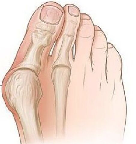
Vallus del Pollice dello del Foat Vallus is the most frequent acquired deformation of the foot, which is characterized by the deviation of the thumbs towards the outside.
The pathology is bilateral nature, mainly affects women over the age of 35.In most cases, the deformation is accompanied by the chronic inflammation of the joint bag and the deforming illegal of the joints of Plusnephalang, pronazional deviations of the first metatarsal bone.
Causes of the disease.Why is it dangerous?
The etiological causes of Vallus Del Pollice dello Della Foot deviations have not yet been completely studied.There are several occurrence theories:
- Vestigial theory.In the middle of the nineteenth century, it was believed that only the models were subject to this deformation due to the use of high -wheeled model shoes, but during the study this pathology was found in men who wore flat shoes.
- The theory of primary muscle weakness - was refuted in a detailed study of the problem.
- The theory of weakness of the binding apparatus and the lack of aponeurosis of the only one - most scientists adhere to this theory.
There are a series of factors that led to the debut of this pathology:
- Excess weight increases the load on the feet.
- Districtly related to age changes in the ligamentary joint system.
- Weekens of Taccod Shoes are more than 5 cm, restricted to the tip.
- The existing deformations of the skeleton (scoliosis, deformation of the femoral joints and knee, the flat feet).
The danger of pathology lies in the fact that over time this deformation leads to the deformation of posture, it is possible to develop dystrophic depths of the spine, hip and knee joints due to the improper distribution of the load during the walk and the lesions to the foot muscles are accompanied by constant pain, which significantly worsens the well -being of a person.
Clinical events
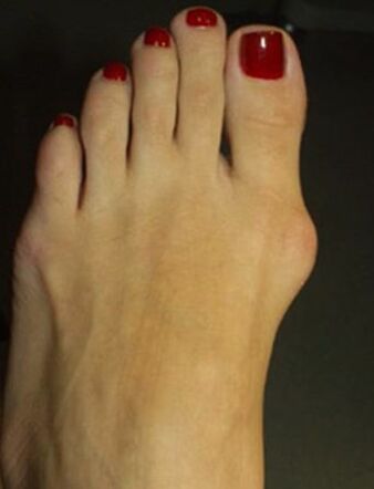
At the beginning of the disease, Valgus deformation does not show itself clinically.Most women are more worried about the appearance of the increase in a foot in the metacarpal-falan joint and the outbreak of the first finger becomes evident.
Over time, pain occurs, first when wearing tight shoes and prolonged walks, and later the pain begins to be constant, sore.
Due to the improper distribution of weight, the limb, from the first and the second finger on the sole, are formed by the seeds, the second finger is raised, strongly extended, the fingers "Malleus" are formed, which is complicated considerably to walk.Due to the deterioration of the blood circulation and the nominee in the front of the foot, arthrosis and chronic bursitis develop.
Diagnostics
- During an objective exam, the classic deformation with deviations of Valgo del Pollice is visible, the distal part of the foot has been expanded.In the projection of the head of the first metatarsal bone, there are signs of hoaxes: hyperemia and skin edema, pain during palpation.
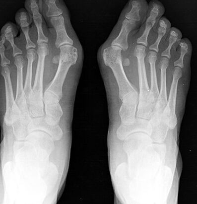
- The X -ray of the foot in two projections.The front projection determines the degree of the thumb, the state of the metacarpal-falan joint, the degree of movement of the sesamoid bone and in the lateral projection the degree of flat feet is displayed and calculated, which often occurs with the deviations of Valgus of the first finger in the foot.
- Plantography.In the printing of the foot, built on paper, a line is traced through the center of the heel and between the fourth and third fingers, the external set of the foot is assessed, according to which the presence of flattening of the foot and its degree is assessed.In these patients, a flattened foot or feet dishes of the 1st degree is more often detected.
Treatment and prevention
Depending on the severity of the process, a conservative or surgical treatment is carried out.
Conservative treatment is performed in the mild phase of the disease, various types of orthopedic sunspasses are used, which are selected individually and bring the deformed finger in the normal position, while the load on the front of the limb is stabilized and distributed.
In the age of children and seniles, the transversal bandage of the distal limb with a pose between the first and the second finger is used.To reduce pain, hot baths are used with sea salt and soda, the radon baths give a good result.
To combat Borsite, anti -local -inflammatory gels are used, compressed with dimexide and lidocaine and are used with pronounced pain, novocaine block and intra -articular glucocorticoid injections.
Surgical treatment is possible at any phase of the disease, it is carried out under local anesthesia, which reduces the list of contraindications for surgery.
In the first phase, the bone excrements are removed on the internal edge of the bone, while it is only possible to reduce the pain of the process, but not prevent the development of the deformation in the future.
With a serious deviation of the finger with the presence of flat feet, an osteotomy is performed at the base of the bone more than the first finger using a bone blade to refuse the finger on the normal position, with the help of a Lavsan ribbon, the transversal ligament of the sole is formed.After surgery, the leg is strictly fixed with a bandage for 4 weeks, it is advisable to use individual orthopedic sunshine during the year.
The prevention of the disease is to use comfortable shoes without high heels, the shoes should sit freely on the leg, a restricted tip should not bring inch discomfort.If there are deformations of the skeleton, it is recommended to undergo preventive exams at the Orthopedist in order to identify and slow down the progression of the deformation of the thumb valley of the foot.
Consequences and complications
A complication of the process is the development of Deichländer or "March" of the foot, which is characterized by acute pain, due to the microocack in the joints of combination of the foot and the trend.
Due to the constant inflammatory process and mechanical damage, there is the risk of developing malignant neoplasms of the bone.